Pyloric stenosis, aka, Infantile hypertrophic pyloric stenosis (IHPS) is a condition affecting infants, characterized by hypertrophy and hyperplasia of the pyloric muscle, leading to gastric outlet obstruction.
- Infants are well at birth. Then, at 3 to 6 weeks of age, the infants present with “projectile” vomiting, potentially leading to dehydration and weight loss.
- More common in first born males
History and examination
- Infants, usually between 2-8 weeks of age
- Risk factors: First-born males(30-40%) , family history, Caucasian ethnicity, bottle feeding, preterm birth, heavy smoker mother
- History of progressive, projectile, non-bilious vomiting, usually after feeding; weight loss and dehydration may be present
| Symptoms | Signs |
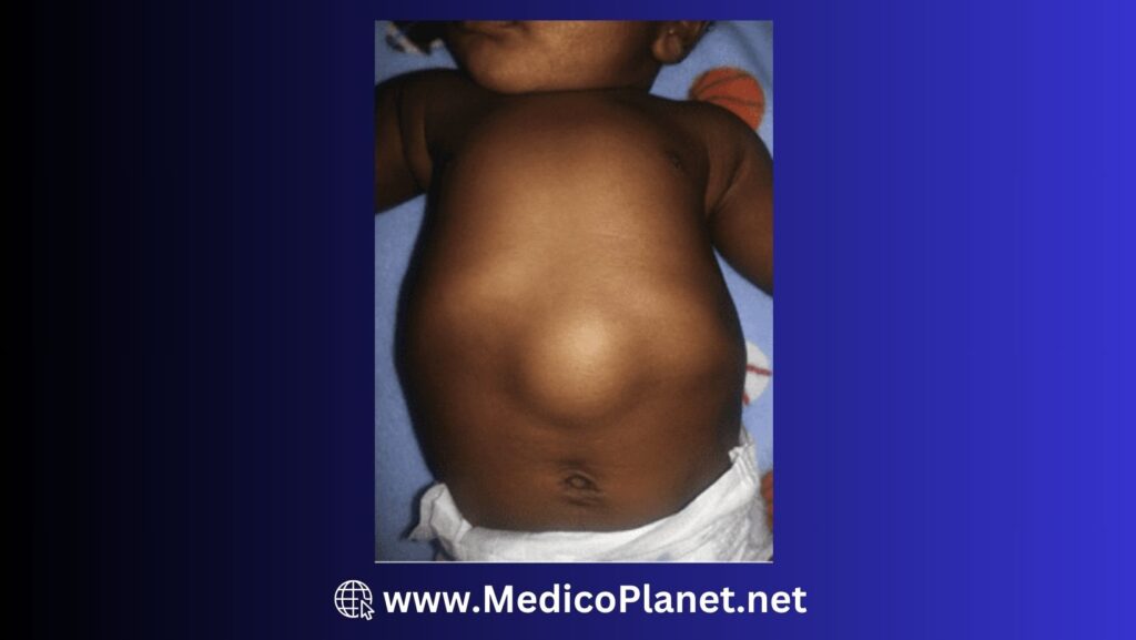
| Projectile vomiting after feeding, non-bilous Persistent hunger Weight loss or poor weight gain | Dehydration (sunken fontanelle, dry mucous membranes, decreased urine output) Visible gastric peristalsis (Peristaltic Abdominal wave) Palpable pyloric(1-2 cm in diameter) “olive” in the right upper quadrant |
Differential diagnosis (D/D) & Complication
D/D:
- Gastroesophageal reflux (Less severe vomiting, no palpable mass)
- Infantile colic (Crying and discomfort, no vomiting)
- Intestinal obstruction (Bilious vomiting, abdominal distension)
- Mid gut volvulus ( first month of life, bilous vomiting)
Complications:
- Dehydration and electrolyte imbalances
- Failure to thrive
Note: In Neonate, vomiting is common. Usually, it is Gastroesophageal reflux in which vomitus is curdy white in consistency, non-projectile, and non-blood stained. In Pyloric stenosis, baby is eager to eat and looks dehydrated and thinly built.
Also, Babies sometimes gulp in air while they’re eating, especially when they drink milk from a bottle. This is also one of the cause of vomiting. In such condition, there will be bloating and excessive passage of gas too. Giving baby too much milk in one go can also cause vomiting.
Treatment for Gastroesophageal reflux and bloated stomach is Burping technique.
Investigation
- Renal function test (to assess dehydration and electrolyte imbalances)
- Hypochloremic, hypokalemic metabolic alkalosis
- Hypernatremia or hyponatremia; both can lead to prerenal renal failure
- ABG
- Abdominal ultrasound (preferred) to visualize pyloric muscle thickness and length; upper gastrointestinal contrast study may also be used.
Admission criteria
- Infants with confirmed pyloric stenosis or severe dehydration requiring fluid resuscitation
Management
Emergency Management: Fluid and electrolyte correction to address dehydration and any imbalances
Medical: No specific medical management for pyloric stenosis
Surgical:
Indication: Confirmed diagnosis of pyloric stenosis
Procedure: Pyloromyotomy (either open or laparoscopic)
Advices
- Monitor for signs of dehydration and electrolyte imbalances post-operatively
- Gradually reintroduce oral feeding after surgery as tolerated
Referral
- Refer to a pediatric surgeon for confirmation of diagnosis and surgical management
References
- Hernanz-Schulman, M. (2003). Infantile hypertrophic pyloric stenosis. Radiology, 227(2), 319-331.
- Panteli, C. (2009). New insights into the pathogenesis of infantile pyloric stenosis. Pediatric Surgery International, 25(12), 1043-1052.
- Le, H. Q., & Baird, R. (2018). Hypertrophic pyloric stenosis: it’s all in the genes! Pediatric Surgery International, 34(4), 383-388.